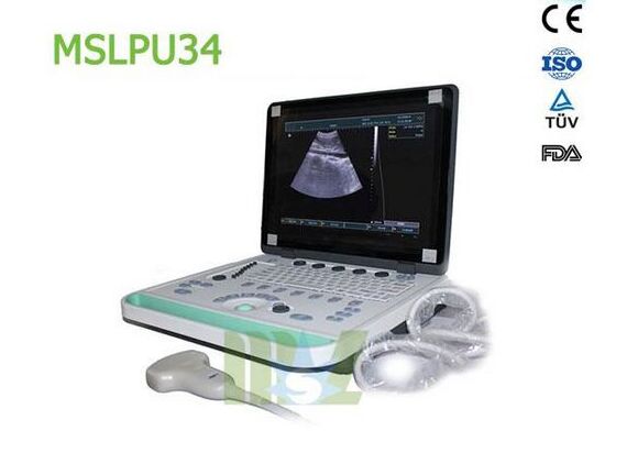
Three-dimensional ultrasound
First talk about Three-Dimensional Ultrasound
A Three-Dimensional Ultrasound belonging of one Color Doppler Ultrasound, Three-Dimensional Ultrasound is a dynamic display. And the superiority of Three-Dimensional Ultrasound including uterine artery, ovarian blood flow sensitivity, display rate; shorten the examination time, to obtain an accurate Doppler spectrum; without filling the bladder, not obese, abdominal scars, intestine inflatable interference; activities by means of the probe tip to find the site of pelvic organ tenderness determine whether pelvic adhesion.
Three-Dimensional Ultrasound imaging surface for obstetric examination, fetal growth can be observed not only the process, but you can check the changes in the placenta, amniotic fluid and umbilical cord. More importantly, it can be used as the primary means of diagnosis of fetal malformations.
Due to the large organizational structure and liquid gray contrast, it can clearly show the three-dimensional shape of a suspicious structure, surface characteristics, spatial relationship to provide three-dimensional images of the fetus within the uterus. Reconstruction include surface imaging, transparent imaging and multidimensional imaging mode.

SecondlyThree-Dimensional Ultrasound features
Increase coronal images based on the two-dimensional image on reservations; three-dimensional positioning, axial adjustment, the three ABC axial plane can be adjusted until the optimum image; three-dimensional imaging, dynamic and intuitive; real-time dynamic observation of the fetal head, the body surface and visceral activities, the image is clear accurate and reliable; cutting function, can keep the image focus; cut out unwanted part of the suspicious sites displaying three-dimensional reconstruction; the rotation function, multifaceted observation; with front and rear, left and right, up and down 360 ° rotation, the image is different orientations comprehensive observation; to the fetus great pictures, record changes in the expression and burn a disc as data retention, aside for a permanent memorial; and to display three-dimensional relationship and adjacent relationship between the different levels of the lesion included.
Four-dimensional ultrasound
Four-Dimensional Color Ultrasonic diagnostic apparatus is the world's most advanced color ultrasound equipment now. “4D” is a “four-dimensional” acronym. The fourth dimension is the time of this vector. For ultrasonic is, 4D ultrasound technology is newly developed technology, but also Canada's exclusive technology winning. 4D ultrasound technology is the use of 3D ultrasound images plus time dimension parameters. This revolutionary technology can obtain real-time three-dimensional images, beyond the limitations of traditional ultrasound. It offers including abdominal, vascular, small parts, obstetrics, gynecology, urology, neonatal and pediatric and other areas of many applications. As a result: the ability to display your unborn baby's real-time dynamic moving images, or other internal organs of the human body in real-time moving image.
Compared with other 3D Ultrasound Diagnostic procedures, 4D Ultrasound so that the doctor can observe real-time dynamic movement of human internal organs.
For example: a doctor can be judged according to fetal movement fetal development, through the observation of three-dimensional planar motion of the needle can improve the accuracy of ultrasound-guided biopsy. So clinicians and doctors Ultrasound can detect and detection of anomalies.
In a word, Four-Dimensional Ultrasound is a complete ultrasound system, which can be used for breast imaging, intervention urology and general medical imaging. As a complete ultrasound equipment Four-dimensional ultrasound used to carry out the following research: Determination of Fetal Age; Analysis of fetal development; Evaluation of multiple births or/and high-risk pregnancies; detect fetal abnormalities; detection of structural abnormalities of the uterus; detect placental abnormalities; detection of abnormal bleeding; detect ectopic pregnancy and other abnormal pregnancy; detection of ovarian cancer, fibroid and placenta Location.
Mothers' 4D Cinema
Moreover, because of the high-definition image quality, Four-Dimensional Color Ultrasonic diagnostic apparatus can achive mother's 4D Cinema. Four-Dimensional Color Ultrasonic diagnostic apparatus can automatically intrauterine fetal shoot “photo” and dynamic video, for many mothers added peace of mind and taste. Which means, for many pregnant mothers, they no longer just feel the baby's breathing and movement, but also can witness their every move and clever show content. More importantly, the four-dimensional ultrasound can be multifaceted, multidimensional observation of fetal growth and development, in order to provide accurate scientific basis for early diagnosis of fetal congenital surface deformities and congenital heart disease. Past B-device can only check the physiological indicators of the fetus, while the four-dimensional ultrasound fetal surface can be checked, such as cleft lip, brain, kidney, heart, skeletal dyspepsia, etc. (requires good fetal position), in order to as soon as possible for treatment. Birth to a smart and healthy baby, and the baby's appearance and actions made into a photo or VCD, so that the baby has the most complete albums when 0 year, which is no longer a fantasy nowadays.
In addition, the four-dimensional color ultrasonic diagnostic apparatus do excellent ergonomic design, radiation rays, electromagnetic waves and other aspects do not exist, what’s more, there is no harmful on human health.


