Sound waves are mechanical vibrations in an elastic medium spread, due to the different ways of vibration and transmission, it can be divided into many types. Of which the most important are longitudinal wave, shear wave, wave and so on. By shock waves in different ways it can be divided into continuous wave and pulsed wave. Medicine for diagnostic ultrasound is a longitudinal wave.

Due to vibration of different frequencies produce different frequencies of sound waves. When the vibration frequency range is 20-20000Hz, the human ear can hear the vibration frequency (number of vibrations per second) in 20000Hz (Hz) or more, the human ear can not hear, this time claiming to produce ultrasound, referred to as ultrasound. But lower than 20HZ human ear can not hear sound waves, then called infra sound, little medical value.
Ultrasound and acoustic vibrations are spread in the elastic medium, it is a mechanical pressure waves. With this launch ultrasonic vibrations to the body, when the ultrasonic encounter two different densities of interface organs or organ and lesions interface, it can be reflected back, take advantage of this situation reflected signals or diseased organ to judge, we called it ultrasonography.
While for Ultrasound, which refers to the causes fluctuations in the human auditory organ, the human ear's hearing threshold range, the vibration frequency of 16kHz-20kHz, exceeding the upper limit of human hearing threshold of sound waves, that is greater than 20kHz sound waves called ultrasound, referred to as ultrasound, clinical ultrasound frequency commonly used in 2-10MHz.
From the angle of the development of ultrasonic diagnostic apparatus, through the A, M-type and B-type ultrasonic diagnostic apparatus, now B-type ultrasonic diagnostic apparatus includes not only the A, M-type functions, also contains a two-dimensional color Doppler imaging, three-dimensional imaging and other functions.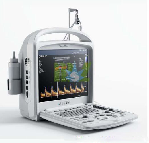
According to differ applications, Ultrasound Classification has been divided in different ranges. First, according to the grade into black and white super, false color and color Doppler ultrasound. Second according to obtain information space is divided into one-dimensional, two-dimensional, three-dimensional, four-dimensional devices. Next according to the image display is divided into A, M-type, B-type, 2B type, B + M type, 4B type, D-type, in addition to A and D, the other belongs to a broad range of B type, is B-Ultrasound. Forth, use is divided into cardiac-specific, dedicated obstetrics and gynecology, abdominal special, dedicated urology, ophthalmology special. And according to shape into a hand-held, laptop or portable, etc.


