Ultrasound Introduction
As we known before, the causing fluctuations in the human auditory organ is called the acoustic sound perception, the human ear's hearing threshold range, the vibration frequency of 16kHz-20kHz, exceeding the upper limit of human hearing threshold of sound waves, that is greater than 20kHz sound waves called ultrasound, referred to as ultrasound, clinical ultrasound frequency commonly used in 2-10MHz.
From the angle of the development of ultrasonic diagnostic apparatus, through the A, M-type and B-type ultrasonic diagnostic apparatus, now B-type ultrasonic diagnostic apparatus includes not only the A, M-type functions, also contains a two-dimensional color Doppler imaging, three-dimensional imaging and other functions.
Ultrasound Classification
· According to the grade into Black and White, False Color and Color Doppler Ultrasound.
· According to obtain information space is divided into One-Dimensional, Two-Dimensional, Three-Dimensional, Four-Dimensional Ultrasound Devices.
· According to the image display is divided into A, M-type, B-type, 2B type, B + M type, 4B type, D-type, in addition to A and D, the other belongs to a broad range of B type, is B-Ultrasound.
· Use is divided into Cardiac-Specific, Dedicated Obstetrics and Gynecology, Abdominal Special, Dedicated Urology, Ophthalmology Special.
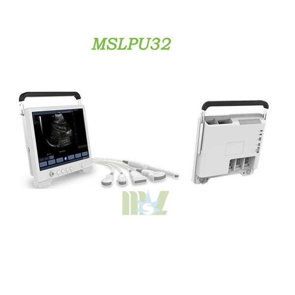
· According to shape into a type of Hand-Held, Laptop, Portable, etc.
B-Mode Ultrasound Diagnosis
B type (brightness mode) ultrasound diagnostics: Genus luminance modulation type, referred to as B-type (B-mode) or B ultrasound, is the echo signal to the spot light and shade, namely gray (gray scale) in the form displayed. Echo signal light spot on the light, the echo signal weak light on the dark point, no echo signal forming dark area, the echo signal from the point, the line to be probed surface structural parts of the two-dimensional tomographic image or a developing section, also known as ultrasonic tomography was like the diagnostic method or section developing method, this image says Sonographic (sonogram).
This method is fast imaging, which can show animal internal organs and diseased section image immediately, which can make static observation (such as the liver, spleen, kidney, uterus, abdominal tumor observation and analysis), but also for the dynamic observation (such as the heart, the fetus was observed). Like using a non-invasive, bloodless “scalpel” to cut layers of organs were observed, which is difficult to achieve other observation methods.
Although the X-ray CT has this ability, but because of its expensive equipment, not widely used. B ultrasound compared with the X-ray CT, not only low prices, but also intuitive and strong for people use in lives, moreover, the technology is simple and easy to master. So in recent years, ultrasound tomography technology to become the most active fertile, the fastest growing a diagnostic technology.


