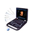
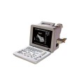
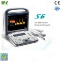
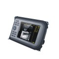
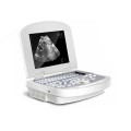
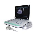
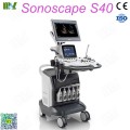
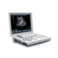
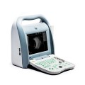
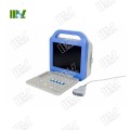
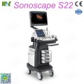
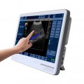
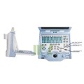
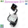
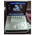
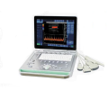
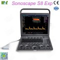
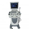
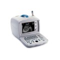
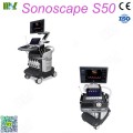
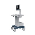
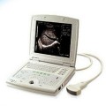
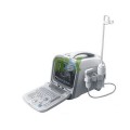
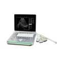
Welcome select our Medical Ultrasound machines MSLCU24, We aim to supply cheaper medical equipments with good service in the same Medical level. And we are ready serve you Specialized Medical Ultrasound machines. Latest new Ultrasound machines, Global Specialized Medical Equipment supplied for different Businesses people by Guangzhou Medsinglong Medical Equipment Co., Ltd.
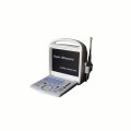
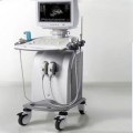
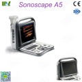
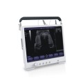
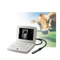
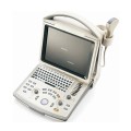
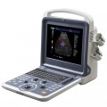
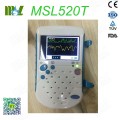
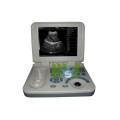
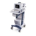
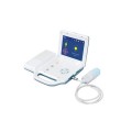
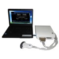
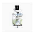
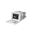
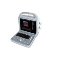
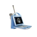
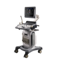
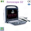
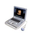
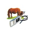
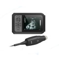
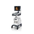
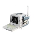
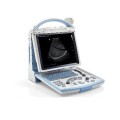
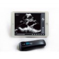
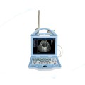
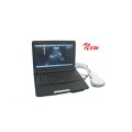
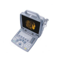
There are Ultrasound machines including: Trolly ultrasound machine, 3D& 4D ultrasound machine, Portable/ Handheld Ultrasound Machines, Home Ultrasound Machine, Veterinary Ultrasound, Color Ultrasound Machine and Digital Ultrasound Machines in differ functions for different person.
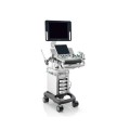
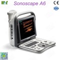
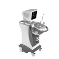
As Professional 4D ultrasound machine manufacturer, Guangzhou Medsinglong Medical Equipment Co., Ltd. supply best price 4D ultrasound machine for sale, MSL 4D Color Doppler Ultrasound Machine/ 4D Baby Ultrasound pregnancy MSLCU24 included.
Product Introduction
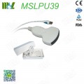
The standard probe showed as below:
TC60A convex array probe TL40A linear array probe
TC10A cavity probe TP16 phased array probe
Briefly principle
* The color Doppler B ultrasound instrument using the probe, make the convert electric excitation signal for ultrasonic signal, sen to the patient’s body, and then reflected the echo signal through the probe and converted to electric signals, processed by the digital circuit. The computer processing system will responsible for the complete emission signal timing and pulse width control, complete echo signal amplifier, dynamic focusing, the TGC (time gain control), the logarithmic compression and signal processing.
* The color Doppler B ultrasound is use the Doppler signal processing, using the technology related to the autocorrelation technique to obtain the blood flow signals after color coding in real-time overlay on the 2 d image, namely the formation of color Doppler ultrasound blood flow images.
The main advantages are:
1) Can quickly visual display two-dimensional plane distribution of the blood flow state;
2) Show the flow direction of the blood;
3) It is convenient to identify the arteries and veins;
4) It’s helpful to understand the nature of the blood flow;
5) It is convenient to understand the speed of the blood flow;
6) Can reliably found shunt and regurgitation;
7) Can make a quantitative analysis for the beam, width, length and area of the blood flow;
Field of Application and Contraindication
Field of Application
The instrument mainly applies in the human body heart, abdominal organs ultrasonic imaging and blood flow movement information collection;
Contraindication
The instrument can not be used in the internal organs examinations like stomach, intestines and lungs which contain the gas. Patients with body burn, scalds or other trauma, cannot be used in the injured part.
Main Functions and Technical Indexes
Operating environment
Operating ambient temperature: + 5℃ ~ + 40℃
Relative humidity: ≤ 80 %
Atmospheric pressure: 700hPa - 1060hPa
Power supply: AC220V ± 22V, 50Hz ± 1Hz
Plug seat shall have independent power supply.
Be far away from strong electric field, strong magnetic field equipment and high voltage equipment
Main Functions
Having a full digital beam forming technology
Scanning mode: Convex array, lumen, high-frequency linear array, phased array;
Dynamic range: 0 ~ 120dB adjustable;
Display mode: B, B / B, M, B / M, CFM, CMF / B, PDI, B / PW, total eight mode;
Application mode: abdomen, gynecology, obstetrics, superficial organ, urologist, heart and user defined model 1- 4, total ten models;
Image mode: digital beam forming, tissue harmonic imaging;
Acoustic output: Mechanical index and thermal index real-time display;
Acoustic power: Step is adjustable, real-time display;
Gray scale: 256 scales;
Depth display: ≥ 250 mm;
B/ D dual-purpose: linear array: B/ PWD; convex array: B/ PWD;
Pseudo color processing: 16 kinds of pseudo color encoding can optional;
Gain adjusts: 8 segments TGC, B/ M/ D/ C is independently adjustable;
TGC curve can show and hide automatically;
Image magnification: picture in picture zoom in and zoom part function;
Image processing:
Edge enhancement: Multilevel adjustable
Frame average: Multilevel adjustable
Line average: Multilevel adjustable
Focus Optimization: Multilevel adjustable
Gray Restrain: Multilevel adjustable
Gamma correction: Multilevel adjustable
Contrast: Adjustable
Brightness: Adjustable
Self-motion optimize function:
Built-in multiple check type, according to different inspection organs, preset best image check condition, reduce the adjusting operation keys;
One-click optimization function:
preset several parameters adjusting focus on a button, a key to realize image fast optimization;
Measurement and calculation:
B mode routine measurement: Distance, circumference, area, volume, angle, ratio, and stenos rate.
M mode routine measurement: Heart rate, time, distance, speed, ratio, etc. Gynecology measurement: Uterus, cervix, endometrial, ovary, follicular.
Obstetrics measurement: EGA, ETD, fetal weight estimation, AFI index, OB report ( including OB tables ).
Cardiology measurement: LV measurement.
Urology measurement: Prostate volume, displacement volume, bladder capacity, and residual urine output.
PW her measurement :
Slice volume measurement, hip joint angle measurement.
Image storage:
Image storage, video storage, cine loop, disk storage capacity ≥ 160G;
Patient data:
Medical record management, report inquiry and printing, image video output (HDD,DVD-RW,USB),built-in ultrasound workstation;
Reporting system: automatic report generation system, and can be full screen characters in both Chinese and English editor;
Output interface: SR323, USB, DICOM interface;
Main Technical Indexes
The performance requirements of gray-scale imaging mode
The color ultrasonic at the gray-scale imaging performance mode should comply with the provisions of the table 3.1
Table 1 At the Gray-scale imaging mode the performance of the probe
|
performance indexes |
probe type and nominal frequency |
|||
|
2.0≤f<4.0 |
2.0≤f<5.0 |
5.0≤f<8.0 |
5.0≤f<10.0 |
|
|
a) probe type and model |
phased array (type TP16A) |
Convex array (type TC60A) |
Cavity (type TC10A) |
Linear array (type TL40A) |
|
b) nominal frequency (MHz) |
3.0 |
3.5 |
6.5 |
7.5 |
|
c) Scan depth (mm) |
≥140 |
≥160 |
≥40 |
≥50 |
|
d) Lateral resolution (mm) |
≤3(depth≤80) ≤4(80<depth≤130) |
≤3(depth≤80) ≤4(80<depth≤130) |
≤2(depth≤30) |
≤2(depth≤40) |
|
e) Axial resolution (mm) |
≤2(depth≤80) |
≤2(depth≤80) ≤3(80<depth≤130) |
≤1(depth≤40) |
≤1(depth≤50) |
|
f) Blind area (mm) |
≤7 |
≤5 |
≤4 |
≤3 |
|
g) Transverse geometry precision (%) |
≤20 |
≤15 |
≤10 |
≤10 |
|
h) Longitudinal geometric location accuracy (%) |
≤10 |
≤10 |
≤5 |
≤5 |
|
i) Slice thickness (mm) |
≤5 |
≤5 |
≤5 |
≤5 |
|
j) Perimeter and area measured deviation (%) |
≤±20 |
≤±20 |
≤±20 |
≤±20 |
|
k) M mode time display error (%) |
≤±10 |
≤±10 |
≤±10 |
≤±10 |
The performance requirements of color Doppler imaging mode
a) The color ultrasonic at the color Doppler imaging mode should comply with the provisions of the table 3.2;
b) Color blood flow image should be essentially coincident with the gray-scale image of pipe’s;
c) Blood flow direction should be able to correctly identify, no aliasing phenomenon;
Table 2 at the color blood flow imaging mode the performance of the probe
|
Doppler model |
Phased array |
Convex array |
Cavity |
Linear array |
|
Investigation depth at Color blood flow model |
≥90mm |
≥100mm |
≥40mm |
≥50mm |
|
Investigation depth at Doppler spectrum model |
≥90mm |
≥100mm |
≥40mm |
≥50mm |
|
Blood flow speed reading error |
≤±15% |
|||
The performance requirements of Doppler spectrum mode
a) The color ultrasonic at the color Doppler spectrum mode should comply with the provisions of the;
b) Blood flow speed reading error should comply with the provisions of the table2.3;
c) Pulse wave Doppler mode sampling area cursor position should be accurate;
Table 3 at the color blood flow imaging mode the performance of the probe
|
Doppler model |
Phased array |
Convex array |
Cavity |
Linear array |
|
Investigation depth at Color blood flow model |
≥90mm |
≥100mm |
≥40mm |
≥50mm |
|
Investigation depth at Doppler spectrum model |
≥90mm |
≥100mm |
≥40mm |
≥50mm |
|
Blood flow speed reading error |
≤±15% |
|||
Displayer: 15 inch LCD color display
Running hours: ≥ 8h;
Input power: ≤ 320V;
Host weight: about 80 kg;
Host appearance size: 950 ×520× 1260 (length × width × height) (mm3).
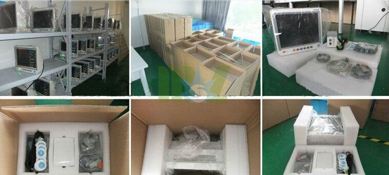
Since the beginning establishment of Guangzhou Medsinglong Medical Equipment Co., Ltd, we have been working in the medical field applications and product innovation. After several years of development, now our products are exported to over 110 countries, the United States, Germany, France, Australia, Turkey, Saudi Arabia, Malaysia and Nigeria included. Furthmore, our company has passed the ISO13485 international quality management system certification, CE certification and RoHS certification.
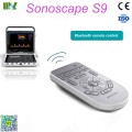
Nowadays, we sell three series ultrasound products including Human ultrasound, Vet animals ultrasound and Ophthalmology ultrasound. Wide ranges of ultrasound such as Notebook B ultrasound, Handheld, 4D ultrasound and other special type of ultrasound products, of which under unique proprietary core technology in the international leading level.
As a developing Medical Devices team, we pursue the goal of "More healthier in technology", and adhere to the idea of "Innovating technology, living healthier". Therefore, we have made a contribution for human health in committing and promoting advanced technologies products for many years.
We are looking forward to cooperating you from differ medical fields around the world, and we firmly believe that we will make you satisfied with our high quality products and good serves.


Welcome to ultrasoundmsl.com, If you have any demand in Ultrasound machine. Please feel free to contact sales@ultrasoundmsl.com
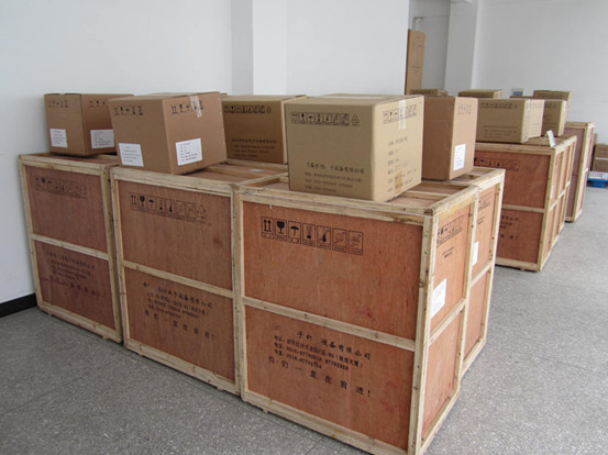
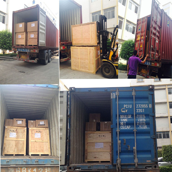
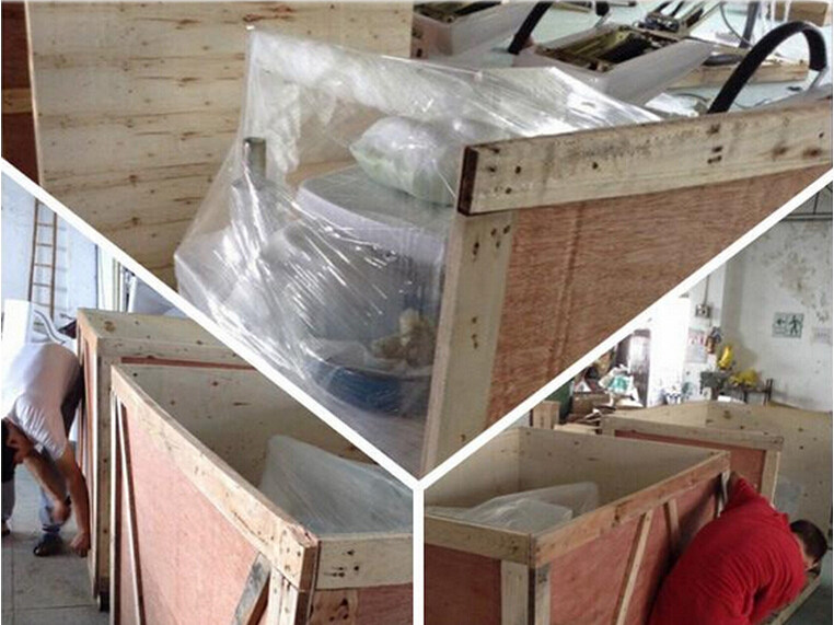
MSL Medical cooperate with DHL, FEDEX, UPS, EMS, TNT, etc. International shipping company, make your goods arrive destination safely and quickly.